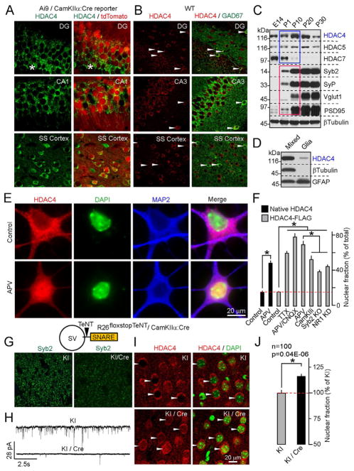
Figure 1. Expression and activity-dependent shuttling of HDAC4 in the forebrain.
A and B, HDAC4 immunoreactivity overlaps with markers of glutamatergic (A) and GABAergic (B, marked by arrows) neurons in the postnatal cortex and hippocampus. In panel (A), asterisks mark the somas of differentiating granule cells.
C, Developmental profile of class IIa HDAC expression in the cortex. Protein extracts were probed by immunoblotting with antibodies to class IIa HDACs, synaptic proteins, and βTubulin (as a loading control).
D, Expression of HDAC4 in mixed cortical cultures and astrocytes.
E, NMDA receptors promote the export of native HDAC4 from the nucleus. Images of control neurons and cells that were treated for 12h with the NMDA receptor blocker, APV, are shown. Scale bar applies to all panels.
F, Quantitative analysis of the subcellular distribution of native and recombinant HDAC4 in wildtype neurons treated with various activity blockers, and Syb2- and NR1-deficient neurons. See Figure S1 for images.
G–J, Nuclear export of HDAC4 is regulated by vesicular release in vivo. G, Loss of Syb2 immunoreactivity in the cortex of R26floxstopTeNT/CamKIIα:Cre mutants (KI/Cre). H, KI/Cre mice have reduced excitatory drive from Entorhinal cortex to DG. Glutamatergic postsynaptic currents were monitored in acute slices from DG granule cells. I and J, KI/Cre mice exhibit accumulation of HDAC4 in the nucleus. Images of brain sections labeled for native HDAC4 (I) and quantifications of HDAC4 localization in the somatosensory cortex (J) are shown. Data from two littermate pairs of KI/Cre and control Cre-negative mice are plotted as Mean±S.E.M. (n=100 neurons per group)

