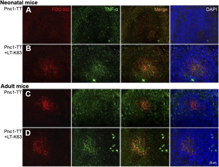
FIGURE 3.
LT-K63 enhanced TNF-α expression that colocalized with FDC-M2 staining in mice immunized with Pnc1-TT as neonates. Spleen sections were stained with FDC-M2 (red), Ab to TNF-α (green), or DAPI to visualize the nuclei (blue) 14 or 10 d after priming of neonatal (A, B) and adult (C, D) mice with 0.5 μg Pnc1-TT (A, C) or 0.5 μg Pnc1-TT+5 μg LT-K63 (B, D), when induction of GCs in spleen peaks in neonatal versus adult mice (14). Seven-micrometer sections were prepared from four different levels, the first started 700 μm into the spleen and the levels were separated by 210 μm. One representative section from each group is shown. Original magnification ×40. Scale bar, 50 μm. Results shown in (A)–(D) are from two experiments for neonatal and adult mice (n = 16 per group), with eight mice per group in each experiment.

