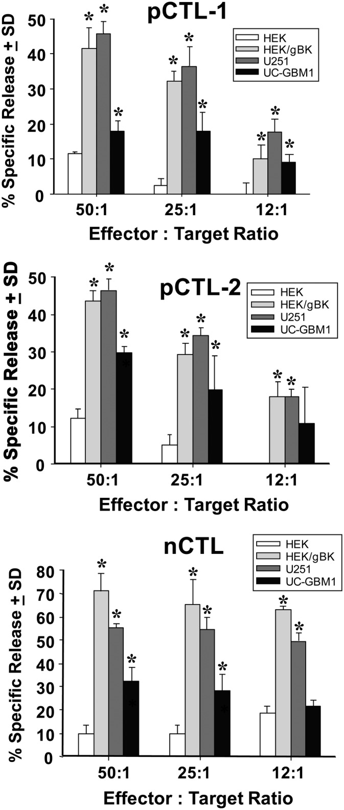
FIGURE 10.

Cytotoxicity experiments with gBK-specific CTL effectors and gBK-expressing and nonexpressing target cells. pCTL-1 and pCTL-2 were generated from GBM patients and nCTLs were stimulated from a normal donor. All CTLs originated from HLA-A0201+ individuals. The DCs isolated from the PBMCs were pulsed with the gBK2 peptide prior to in vitro DC maturation. Autologous T cells were then stimulated and further expanded. The target cells included the U251 glioma cell line, early passage (P2) UC-GBM1 glioma cells (autologous to the pCTL-1), and HLA-0201+ HEK cells that were either nontransfected or transfected with the gBK plasmid 48 h prior to the assay. The gBK profile of the HEK cells is shown in Fig. 3D. All test symbols represent data that were done in quadruplicate cultures. Top panel, Results using the pCTL-1; middle panel, data generated with the pCTL-2; bottom panel, results using the nCTLs. *p < 0.05 from the nontransfected HEK control values.
