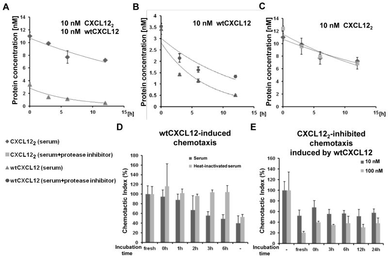
Figure 4. Mouse serum quickly diminishes wtCXCL12 function whereas CXCL122 remains active for at least 24 hours.
(A) Degradation of 10 nM wtCXCL12 and CXCL122 were monitored by ELISA in the presence of mouse serum (n=3). Representative of 2 independent experiments with similar results. (B) WtCXCL12 (initial starting concentration, 10 nM) degradation monitored in the presence of a protease inhibitor cocktail (cOmplete, Mini, EDTA-free (Roche); n=3). (C) Degradation of CXCL122 (initial starting concentration, 10 nM) was monitored in the presence of protease inhibitors. Negative control (labeled “−”) contained no chemokine but contained 1% mouse serum (n=3). (D) WtCXCL12-induced chemotaxis as a function of time the chemokine is incubated with 90% mouse serum (n=3). (E) Inhibition of 10 nM wtCXCL12-induced migration by either 10 nM or 100 nM CXCL122 incubated with 90% mouse serum for indicated time. Chemotaxis assays were performed using THP-1 monocyte cells (n=3). Negative control (labeled “−”) contained no CXCL122 but contained 10 nM wtCXCL12 and 1% mouse serum. As a positive control (labeled “fresh”), the indicated chemokines were incubated only with 1% mouse serum, which did not result in degradation (data not shown).

