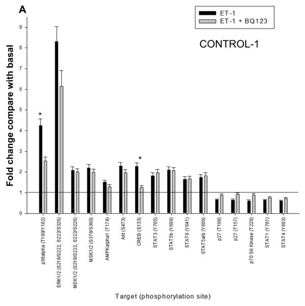
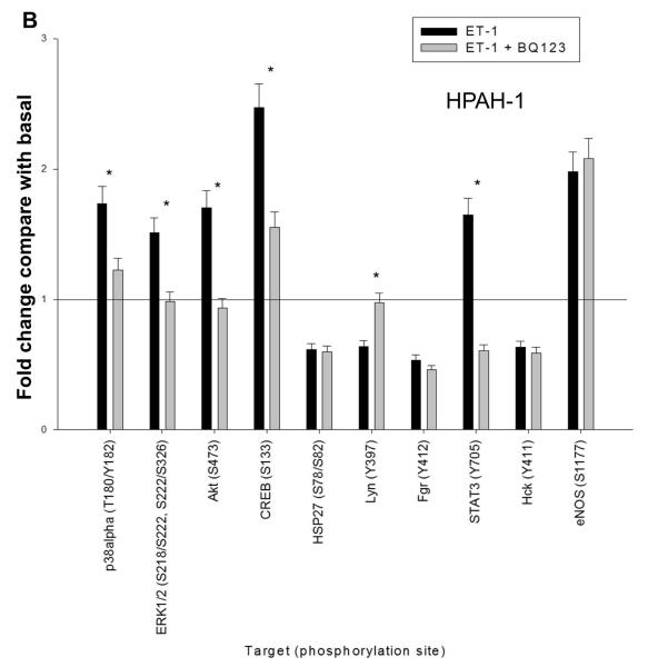
Figure 7. ET-1 mediated phosphorylation events in CONTROL-1 and HPAH-1 PASMC.
The human phospho-antibody array detected phosphorylated proteins in untreated and treated cell lysates from donor control CONTROL-1 (A) and BMPR2 deletion HPAH-1 PASMC (B). The cells were either left untreated or treated with 10 nM ET-1 for 5 minutes with or without 1 uM BQ123. Profiles were created by quantifying the mean spot pixel densities normalized by positive control dots. Array signals from scanned X-ray film images were analyzed using image analysis software NIH ImageJ. The effect of ET-1 or ET1 + BQ123 was presented as fold change compared with the basal level of untreated sample. Only the targets with the fold change greater than 1.5 or less than 0.67 upon ET-1 stimulation were presented. The statistical analysis was performed using t-test. * p < 0.05 compared with untreated basal.


