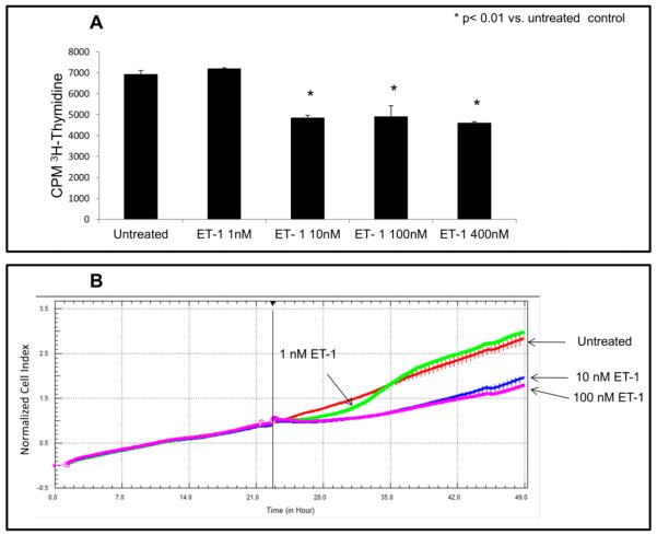
Figure 3. Effects of ET-1 dosage on growth inhibition in CHO ETB cells.
(A) 3H-thymidine incorporation was performed as previously described on CHO ETB cells but with different ET-1 concentrations (0, 1, 10, 100, and 400 nM). Cells were pulsed with [3H]-thymidine during the interval of 4-6 hours after ET-1 addition. Bar height indicates the CPM average and the error bars are the standard deviation. *p< 0.01 vs. untreated control. (B) Growth curve of CHO ETB cells treated with different ET-1 concentrations (0, 1, 10, and 100 nM) generated by real-time cell impedance measurements. ET-1 was added 24 hours after cells were seeded. To offset any small differences in cell growth before ET-1 addition, cell index values were normalized in the graph after ET-1addition. All points in the curves represent the normalized cell index average and the error bars are the standard deviation.

