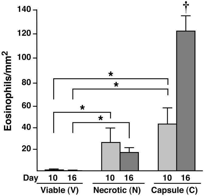
Figure 3. Eosinophils differentially accumulate within the necrotic and capsule regions of B16-F10 melanoma cell-derived tumors.
Quantitative assessments of eosinophil density (i.e., eosinophils/mm2) were derived from serial sections of entire tumors. Eosinophils were counted within the necrotic, viable, and capsule regions from 4μm sections taken every 100μm through each tumor (n=10 mice/group) and expressed as a function of the region’s area. The data presented represent mean averages ± SEM. All evaluations were performed in duplicate as independent observer-blinded assessments. *P<0.05; †, P<0.05 relative to all other groups.

