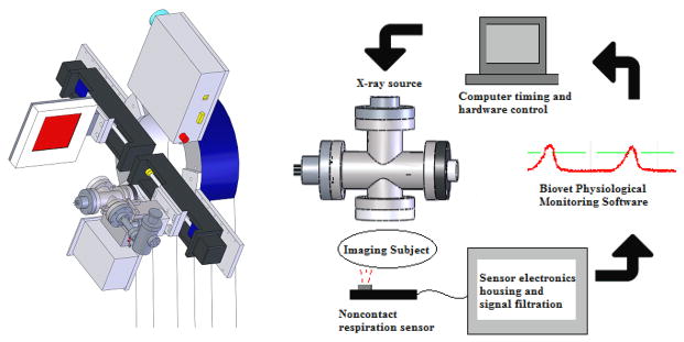
Figure 3.
The custom-built CNT cone beam micro-CT used in this study (left) consists of a compact field-emission x-ray source and flat panel detector mounted adjacent to one another on a rotating gantry. The sensor is integrated into the complete hardware of the micro-CT system as in the flowchart above (right). The sensor’s output is fed into Biovet and the computer’s timing program, so that appropriate physiological gating can be achieved with the x-ray source

