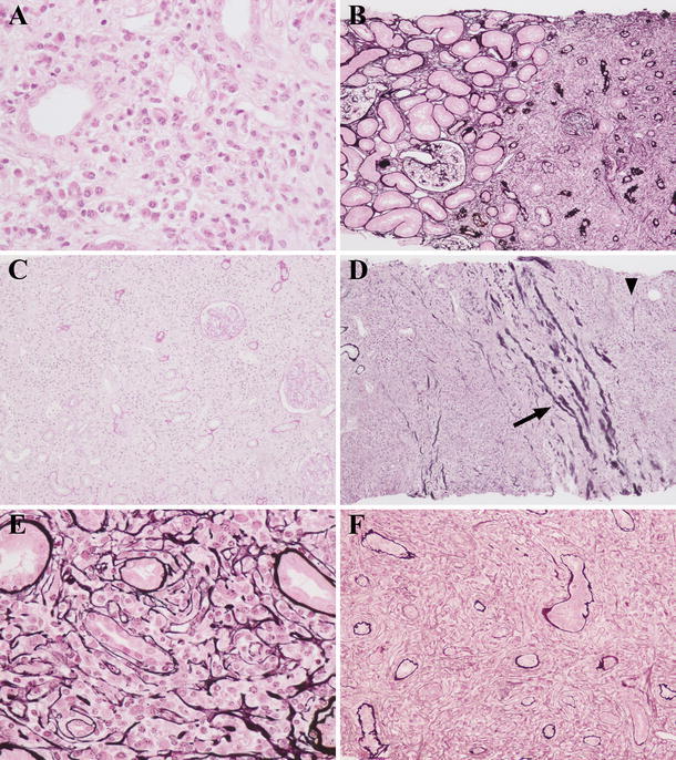
Fig. 3.

Light microscopy findings of the renal interstitium before corticosteroid therapy. Severe lymphoplasmacytic infiltration with tubular atrophy was observed (a patient 6, b patient 4, c patient 6). Interstitial lesions were often focal, and the borderline between lesion and nonlesion was fairly clear (b). In two patients with severe renal dysfunction, interstitial lesion was diffuse (c). Inflammatory lesion beyond the renal capsule was detected (d patient 6, arrow shows the renal capsule, and arrowhead shows inflammation beyond the renal capsule). A characteristic fibrosis that appeared to surround infiltrating cells was observed (e patient 4, f patient 5) [a Hematoxylin and eosin (H&E) staining ×400; b, d periodic acid-methenamine-silver (PAM)-H&E staining ×100; c periodic acid-Schiff (PAS) staining ×100; e, f PAM-H&E staining ×400]
