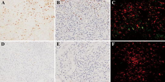
Fig. 5.

Changes in immunoglobulin (Ig)G4-positive plasma cell, Foxp3+ cell, and CD4+CD25+ cell infiltration before (a–c) and 1 month after (d–f) corticosteroid therapy in patient 6. Compared with the pretreatment specimens (a), the number of IgG4-positive plasma cells in the lesions was markedly diminished in the posttreatment specimens (d). In the same way, compared with the pretreatment specimens (b), the number of Foxp3+ cells obviously decreased in the posttreatment specimens (e). Some CD4+CD25+ cells in the lesions were observed before treatment (c), whereas almost none were detected after therapy (f) [a, d IgG4 ×200, b, e Foxp3 ×400, c, f CD4 (red) and CD25 (green) ×400]
