. 2012 Mar;2(1):57–58. doi: 10.3978/j.issn.2223-4292.2012.01.01

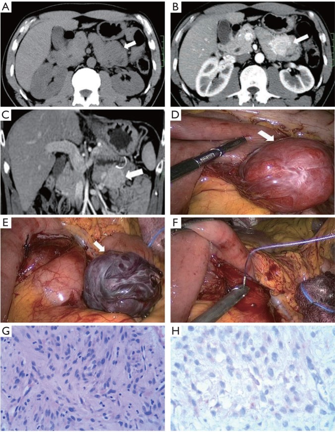
Figure 1. A. Transversal plain CT scan depicts a homogeneous iso-attenuation mass (arrow), but it could not be differentiated from crimpy bowel loops; B. Transversal contrast-enhanced CT demonstrates a homogeneous avidly enhanced mass (arrow); C. Coronal multi-planar reformation of contrast-enhanced CT shows the mass is well-defined (arrow). The musical layer covering the mass (curve arrow) is intact and the fat plane between the mass and surrounding tissue is preserved; D. Laparoscopic photograph shows an exophytic mass with intact capsule; E. Laparoscopic photograph show the mass was completely excised; F. Laparoscopic photograph shows the wound was sewed; G. Photomicrograph (original magnification, ×100; hematoxylin-eosin stain) of the tumor shows spindle cells with pale eosinophilic cytoplasm; H. Immunohistochemistry demonstrates positive staining for CD-117 (original magnification, ×200).

Figure 1.