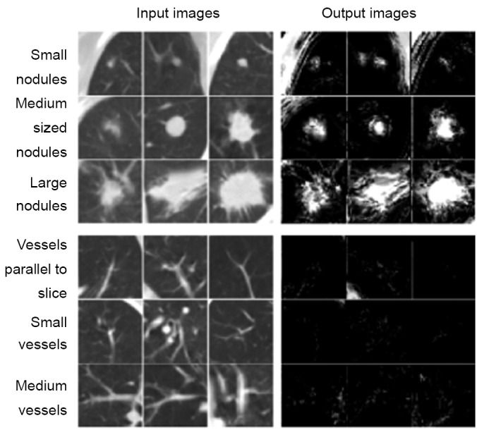
Figure 3.

Enhancement of lung nodules and suppression of FPs (i.e., lung vessels) by use of MTANNs for FP reduction. Once lung nodules are enhanced, and FPs are suppressed, FPs can be distinguished from lung nodules by use of scores obtained from the output images
