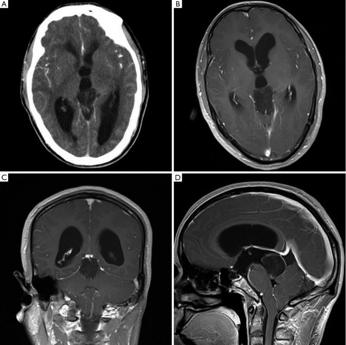
Figure 1.
A. CT shows ventriculomegaly due to a well-defined hypodense lesion in the pineal gland and the lesion showed faint heterogeneous contrast enhancem ent. No calcification was noted; B. MRI confirms a well-defined 3.2 cm × 2.2 cm× 3.0 cm solid-cystic mass in the pineal gland. Contrast enhancement shows mild dotted enhancement inside the mass; C. Coronal MR image shows the mass involved the tectum; D. Sagittal MR image of the lesion shows ring-shaped enhancement and the lesion involved the posterior third ventricle and extended to the coping of the fourth ventricle, but without ventriculomegaly of the fourth ventricle

