Fig. 2.
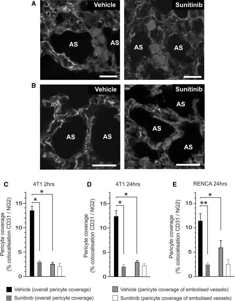
Reduced pericyte coverage in the lung microvasculature is associated with enhanced seeding of metastasis. Balb/c mice were pre-treated with vehicle (veh) or 120 mg/kg/day sunitinib (sun) every day for 7 days, followed by intravenous injection of fluorescently tagged 4T1-luc or RENCA-luc tumour cells. Lungs were harvested at 2 or 24 h post-injection of tumour cells and sections were stained for the vessel marker CD31 and the pericyte marker NG2. a Representative images of 4T1 tumour cell emboli trapped within the lung vasculature at 24 h post-injection of tumour cells in mice pre-treated with vehicle or sunitinib as indicated, tumour cells (blue), CD31 (red) and NG2 (green). b Representative images of pericyte coverage within the lung vasculature at 24 h post-injection of 4T1 tumour cells in mice pre-treated with vehicle or sunitinib as indicated, CD31 (red), NG2 (green) and DAPI (blue). c, d Quantification of overall pericyte coverage in the lung vasculature and pericyte coverage only in vessels containing 4T1 tumour cell emboli. Data are shown at 2 h (c) and 24 h (d) post-injection of tumour cells in mice pre-treated with vehicle or sunitinib. Bar graph shows percentage colocalisation between CD31 and NG2 ± SEM. * P = 0.0001, n = 8 mice per treatment group. e Quantification of overall pericyte coverage in the lung vasculature and pericyte coverage only in vessels containing RENCA tumour cell emboli. Data are shown at 24 h post-injection of tumour cells in mice pre-treated with vehicle or sunitinib. Bar graph shows percentage colocalisation between CD31 and NG2 ± SEM. * P = 0.03, ** P = 0.0001. n = 8 mice per treatment group. Scale bar = 25 μM, AS = alveolar space. (Color figure online)

