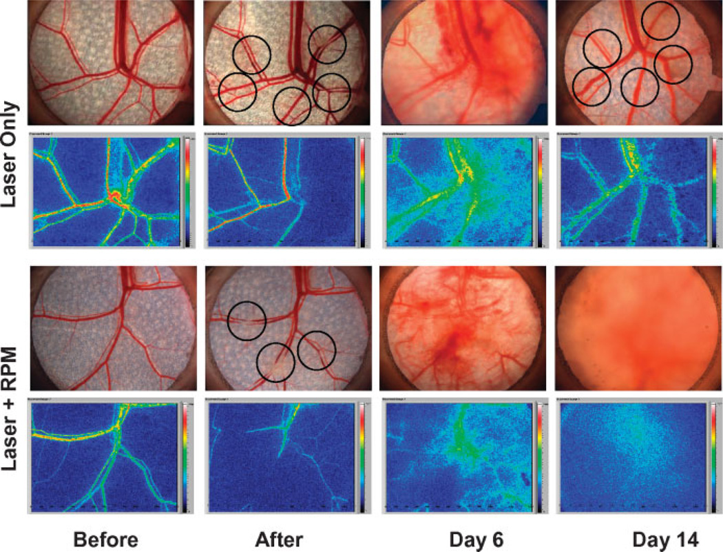
Fig. 3.
Digital photographs of the subdermal side of the RWCM before and immediately after laser exposure, and 6 and 14 days later with or without topical RPM treatment. Circles in “After” panel indicate sites of laser irradiation. Images on Day 6 “Laser Only” panel indicate early signs of blood vessel revascularization. Images on Day 14 “Laser Only” panel indicate complete blood vessel revascularization. Laser speckle blood flow images show changes consistent with complete reperfusion of these vessels. Images on Day 14 “Laser + RPM” panel show no revascularization and reperfusion 14 days after laser irradiation and topical RPM treatment.

