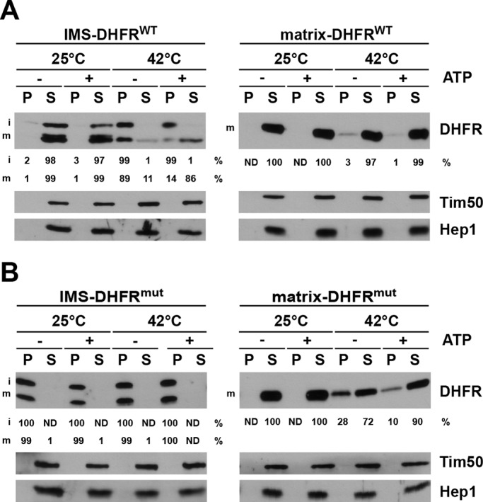
FIGURE 2:

Aggregation of wild-type and mutant DHFR constructs in IMS and matrix. (A and B) Isolated mitochondria were incubated under conditions to increase or decrease the mitochondrial ATP levels and then exposed to 25°C or 42°C for 3 min. Mitochondria were then solubilized with Triton X-100–containing buffer and soluble (S) and aggregate (P, pellet) fractions separated by centrifugation and analyzed by SDS–PAGE and immunodecoration using the indicated antibodies. The DHFR signals were quantified in supernatant and pellet fractions and expressed as percentages of total. ND, not detectable.
