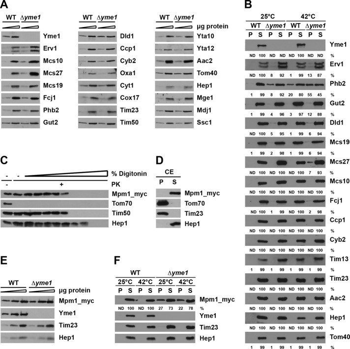
FIGURE 5:
Effect of Yme1 on aggregation of mitochondrial proteins. (A) Mitochondria (5 and 15 μg) were analyzed by SDS–PAGE and immunodecoration with the indicated antibodies. (B) Mitochondria were preincubated for 3 min at 25 or 42°C and solubilized with Triton X-100–containing buffer, and soluble (S) and aggregate (P, pellet) fractions were separated by centrifugation. Samples were analyzed by SDS–PAGE followed by immunodecoration with the indicated antibodies. The DHFR signals were quantified in supernatant and pellet fractions and expressed as percentages of total. ND, not detectable. (C and D) Submitochondrial localization of Mpm1. Mitochondria harboring a myc-tagged version of Mpm1 were subjected to (C) digitonin fractionation, as described in Figure 1, and (D) carbonate extraction (CE). Samples were analyzed by SDS–PAGE and immunodecoration with the indicated antibodies. (E) Steady-state levels of Mpm1 in wild-type and ∆yme1 strain. Mitochondria (5, 15, and 30 μg) were analyzed as in (A). (F) Aggregation of Mpm1 in ∆yme1 cells. Isolated mitochondria were analyzed as in (B).

