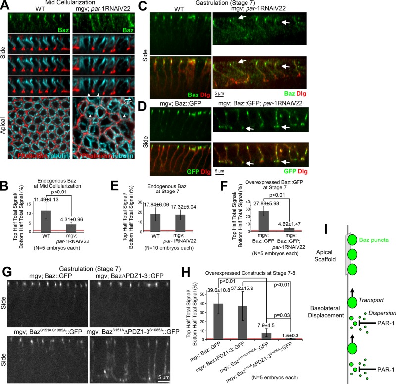
FIGURE 4:
PAR-1 phosphorylation contributes redundantly to Baz polarization. (A) Fixed midcellularization embryos. In WT, Baz localizes apicolaterally, phalloidin stains overall furrows and actin-rich furrow canals, and apical-basal microtubule bundles form baskets around nuclei. With maternal expression of par-1 shRNA (Valium22 line) with mgv, Baz mislocalizes basally and furrows are lost sporadically (arrowheads), but microtubule baskets are present. (B) Total fluorescence intensity ratios of endogenous Baz puncta in the top vs. bottom halves of lateral membranes. (C, D) Fixed stage 7 embryos. (C) With and without PAR-1 RNAi, endogenous Baz is apicolaterally enriched despite PAR-1 RNAi morphological defects. Dlg shows basolateral membranes. (D) Insertion of UAS-Baz::GFP at attp40 (chromosome 2) allowed maternal coexpression with par-1 shRNA (Valium22 line) at attp2 (chromosome 3; other Baz constructs at attp2). With coexpression, Baz::GFP mislocalized basally, accumulating at the base of lateral membranes (arrows). (E, F) Total fluorescence intensity ratios of puncta in the top vs. bottom halves of cells. (G) Fixed stage 7 embryo side views with Baz::GFP, Baz∆PDZ1-3::GFP, BazS151AS1085A::GFP, and BazS151A∆PDZ1-3S1085A::GFP expressed maternally with mgv. (H) Total fluorescence intensity ratios of protein puncta in top vs. bottom halves of cells. Red lines show 1:1 top:bottom ratios. (I) Model of Baz polarization.

