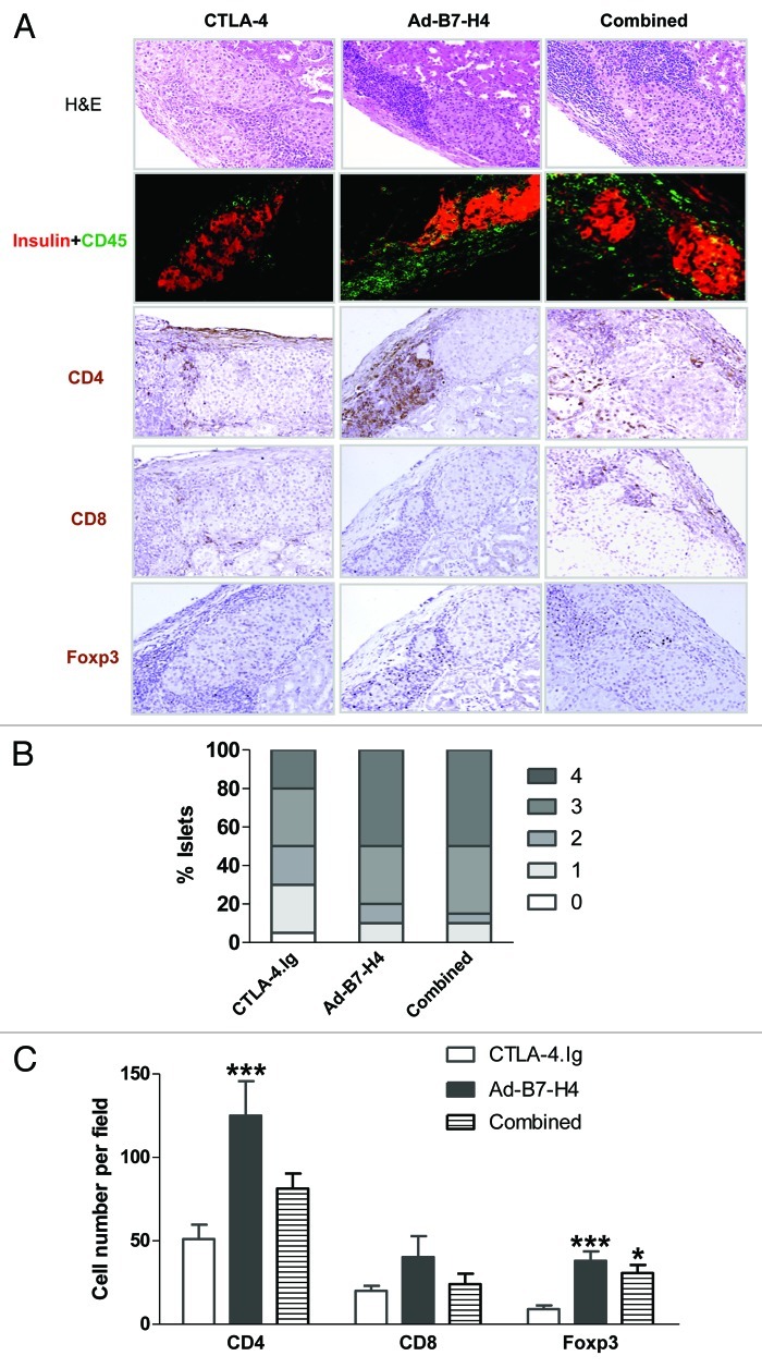
Figure 7. Allografts from long-term surviving recipients treated with CTLA-4.Ig and/or Ad-B7-H4 exhibit distinct expression patterns. β-cell function, infiltrates and subsets of infiltrates were assessed from long-term surviving mice 90 d post-transplant. (A) Representative sections were stained for H&E, CD45 plus insulin, CD4, CD8, and Foxp3 from each treatment group. (B) Isletitis was scored according to staining for H&E, CD45 plus insulin. Sections were randomly selected and blindly scored using four to eight animals per group. Isletitis was graded by Yoon’s method (see Fig. 6). (C) Expression of CD4, CD8, and Foxp3 was calculated according to the numbers of positive-staining cells per field, and the field was randomly selected and blindly scored. Treatment with CTLA-4.Ig exhibited minimal infiltrates (p < 0.001 vs. Ad-B7-H4 by ANOVA). A high number of Foxp3+ cells was found in Ad-B7-H4 and combined groups (p < 0.001 vs. CTLA-4.Ig by ANOVA). n = 6–10 islet allografts per group.

An official website of the United States government
Here's how you know
Official websites use .gov
A
.gov website belongs to an official
government organization in the United States.
Secure .gov websites use HTTPS
A lock (
) or https:// means you've safely
connected to the .gov website. Share sensitive
information only on official, secure websites.
