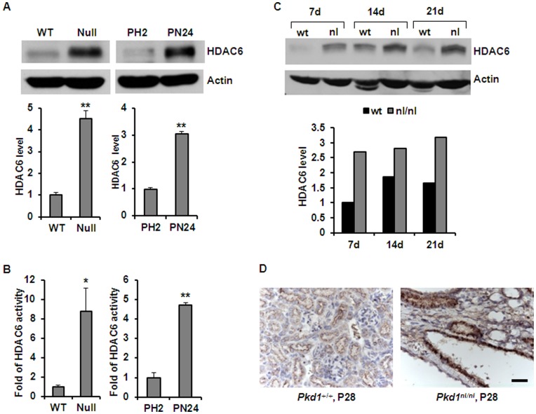
Figure 1. HDAC6 was up-regulated in Pkd1 mutant renal epithelial cells and kidney tissues.
(A) Western blotting analysis of the expression of HDAC6 in Pkd1 wild-type (WT) and Pkd1null/null (Null) MEK cells as well as in postnatal Pkd1 heterozygous PH2 cells and homozygous PN24 cells. The expression of HDAC6 in the above cells was quantified from three independent immunoblots and was standardized to actin. ** p < 0.01. (B) The activity of HDAC6 in Pkd1 wild-type (WT) and Pkd1null/null (Null) MEK cells as well as in postnatal Pkd1 heterozygous PH2 cells and homozygous PN24 cells. The activity of HDAC6 was increased about 7.8 fold in Pkd1null/null (Null) MEK cells and 4.8 fold in postnatal Pkd1 homozygous PN24 cells compared with that in Pkd1 wild type MEK cells and postnatal Pkd1 heterozygous PH2 cells, respectively. The HDAC6 activity assay was performed in three independent experiments. * p < 0.05, ** p < 0.01. (C) Western blotting analysis of the expression of HDAC6 in Pkd1 wild type and Pkd1 hypomorphic homozygous (Pkd1nl/nl) kidney tissues collected at postnatal days 7, 14, and 21. The relative expression levels of HDAC6 in different kidneys were standardized to actin and were presented in the lower panel. (D) Immunohistochemistry of HDAC6 in kidney sections from Pkd1 wild type and Pkd1nl/nl mice harvested at postnatal day 28. Scale bar, 20 µm.

