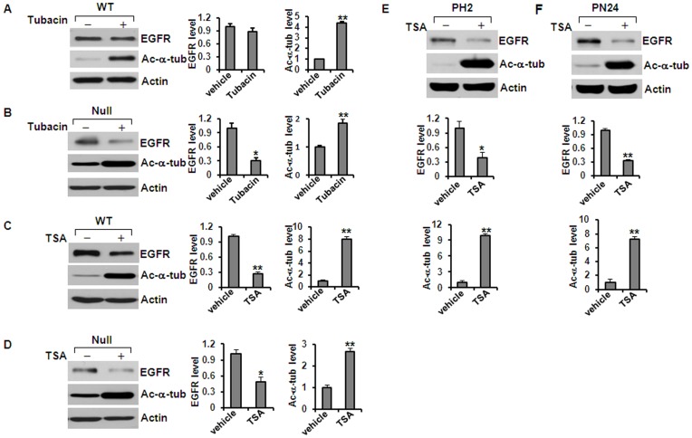
Figure 2. Inhibition of HDAC6 activity decreased the levels of EGFR and increased acetyl-α-tubulin in renal epithelial cells.
(A) and (B) Western blotting analysis of the expression of EGFR and acetyl-α-tubulin in Pkd1 wild-type (WT) MEK cells (A) and Pkd1null/null (Null) MEK cells (B) treated with or without tubacin. Tubacin (5 µM) treatment deceased the levels of EGFR in Pkd1 mutant MEK cells, but increased the acetyl-α-tubulin in both wild-type and mutant MEK cells. (C) and (D) Western blotting analysis of the expression of EGFR and acetyl-α-tubulin in Pkd1 wild-type (WT) MEK cells (C) and Pkd1null/null (Null) MEK cells (D) treated with or without TSA (100 ng/ml). TSA treatment decreased the levels of EGFR and increased the acetyl-α-tubulin in Pkd1null/null (Null) MEK cells. (E) and (F) TSA (200 ng/ml) has the same effect on the expression of EGFR and acetyl-α-tubulin in postnatal Pkd1 heterozygous PH2 and homozygous PN24 cells. * p < 0.05. ** p < 0.01.

