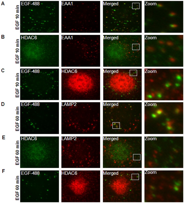
Figure 4. HDAC6 associated with the endosomal compartments in mIMCD3 cells.
(A) mIMCD3 cells were serum-starved overnight and then treated with 80 ng/ml EGF-Alexa Fluor 488 for 10 min. After that, cells treated with 0.005% saponin were stained with anti EEA1 antibody. Image in zoom shows the example of early endosomes stained with EEA1 (red) colocalized with EGFR (green). (B) Serum-starved mIMCD3 cells were treated with 100 ng/ml EGF for 10 min and then stained with anti-HDAC6 and EEA1 antibodies. Image in zoom shows the example of early endosomes stained with EEA1 (red) colocalized with HDAC6 (green). (C) mIMCD3 cells were treated and processed as in (A) except using anti-HDAC6 antibody. Image in zoom shows the colocalization of EGFR (green) with HDAC6 (red). (D) mIMCD cells were treated with 80 ng/ml EGF-Alexa Fluor 488 for 60 min and then stained with anti-LAMP2 antibody. Image in zoom shows the colocalization of EGFR (green) with later endosomes stained with LAMP2 (red). (E) mIMCD3 cells were treated with 100 ng/ml EGF for 60 min and then stained with anti-HDAC6 and anti-LAMP2 antibodies. Image in zoom shows the example of later endosomes stained with LAMP2 (red) colocalized with HDAC6 (green). (F) mIMCD3 cells were treated and processed as in (D) except using anti-HDAC6 antibody. Image in zoom shows the colocalization of EGFR (green) with HDAC6 (red).

