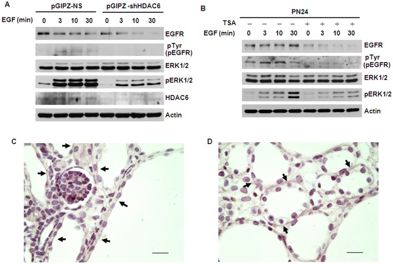
Figure 10. Inhibition of HDAC6 activity decreased the phosphorylation of ERK1/2 and normalized the localization of EGFR from apical to basolateral.
(A) Knockdown HDAC6 with shRNA increases the degradation of EGFR and decreases the phosphorylation of ERK1/2. Serum-starved PN24 cells transduced with lentiviral plasmid pGIPZ-shHDAC6 or control empty vector pGIPZ-NS, were pretreated with 10 µg/ml cycloheximide for 2 hours and then stimulated with 100 ng/ml EGF for indicated times. (B) Inhibition of HDAC6 activity with TSA decreases the activation of EGFR and ERK1/2 . PN24 cells were pretreated with 100 ng/ml TSA for 12 hours and then starved for another 12 hours with TSA. After that, the cells were stimulated with 100 ng/ml EGF in the presence of 10 µg/ml cycloheximide and TSA for indicated times. (C) and (D) Treatment with TSA normalized the mislocalized EGFR from apical to basolateral in cystic epithelia in postnatal day 7 (P7) kidneys from Pkd1flox/flox:ksp-Cre pups. Immunohistochemical staining of EGFR in P7 kidney sections from Pkd1flox/flox:ksp-Cre neonates treated with DMSO (C) or TSA (D). Arrows pointed to apical (C) and basolateral (D) regions, respectively. Scale bar, 20 µm.

