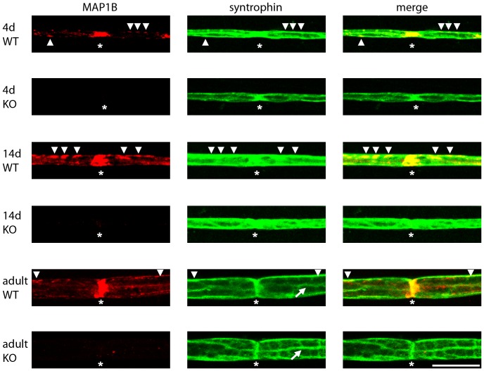
Figure 4. MAP1B and syntrophin co-localize at the nodes of Ranvier and the abaxonal Schwann cell membrane.
Sciatic nerves were prepared from 4-day (4d) or 14-day (14d) old or adult (adult) wild-type (WT) and MAP1B−/− (KO) mice. Individual myelinated axons were isolated and stained for MAP1B (antibody anti-HC750) or syntrophin (pan syntrophin antibody anti-syn1351) as indicated. The pictures represent projections of confocal Z-stacks. The staining for MAP1B in postnatal and adult Schwann cells is specific as it is absent in Schwann cells of MAP1B−/− mice. At all ages MAP1B was found to be concentrated at the nodes of Ranvier (asterisks). It also localized at the abaxonal membrane (arrow heads), particularly strong at postnatal day 14. Syntrophin was also found at nodes of Ranvier and the abaxonal membrane. In the adult, it was found to be localized to Cajal bands (arrows) in agreement with previous results [44]. Co-localization of MAP1B and syntrophin was most prominent at the nodes of Ranvier and partial co-localization was found at the abaxonal membrane (arrow heads). Scale bar, 20 µm.

