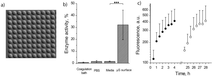
Figure 2. Visualization and initial characterization of µS PVA hydrogels as matrices for SMEPT.
(a) Differential interference contrast microscopy image of the surface adhered µS PVA hydrogels (2 µm cubic structures). (b) Levels of β-Glu enzymatic activity revealed by polymer coagulation bath, PBS washing solution, cell culture media washing solution, and final surface adhered µS PVA hydrogels. Enzyme quantification was performed using FdG, 30 min reaction time and a standard curve obtained for the activity of β-Glu in PBS. (C) Experimental data for conversion of FdG into fluorescent product using µS PVA hydrogels and initiating conversion via administration of FdG at time points t = 0 (solid circles) and t = 24 h (open circles). Experimental conditions: [FdG]: 2,5 µg/mL; 1 g/L β-Glu in the polymer solution used for the production of hydrogels.

