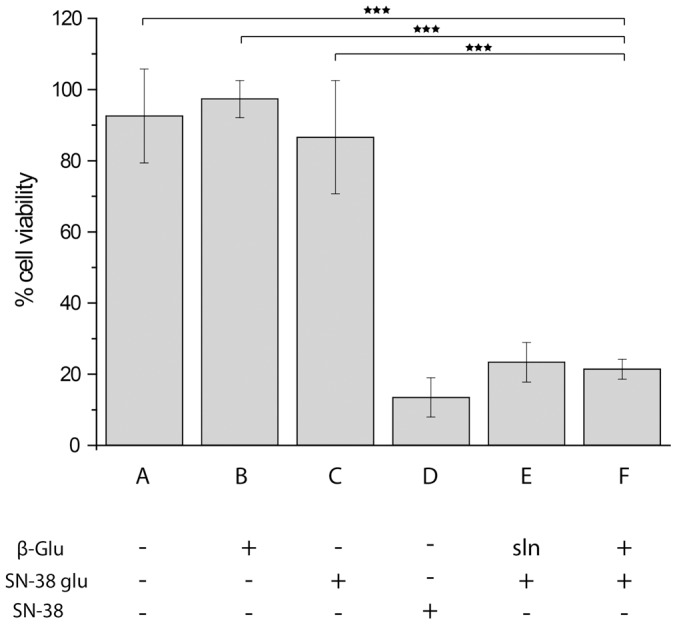
Figure 7. Viability of HepG2 cells cultured on µS PVA thin films as quantified using Presto Blue viability assay and expressed relative to viability of these cells on tissue culture polystyrene at a matched initial cell seeding density.

The cells were cultured on (A) pristine µS PVA hydrogels; (B) µS PVA thin films equipped with β-Glu; (C) enzyme-free µS PVA hydrogels in the presence of 1 µM SN-38 glucuronide; (D) enzyme-free µS PVA hydrogels, 1 µM SN-38; (E) µS PVA hydrogels in the presence of 1 µM SN-38 glucuronide and β-Glu added to the cell media (solution based enzyme prodrug therapy); (F) SMEPT conditions, i.e. β-Glu equipped µS PVA thin films in the presence of 1 µM SN-38 glucuronide. Presented results (mean ± st.dev.) are average over at least 3 independent experiments, 3 replicates each.
