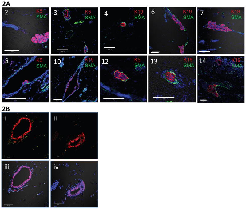
Figure 2.
Staining techniques were used to confirm the presence of primary human cells within the transplanted mouse mammary glands. 2A: Numbers correspond to the case numbers which successfully transplanted. Representative immunofluorescent staining of human CK 5 (K5) or 19 (K19), smooth muscle actin (SMA), and DNA. K5 and K19 are conjugated to Alexa-fluor 594, shown in red, and SMA is conjugated to Alexa-fluor 488, shown in green. Nuclei are counterstained with DAPI. 2B: Representation fluorescent in situ hybridization (FISH) analysis of a MIND xenograft using probes labeled for mouse DNA, human DNA, and DAPI. The human DNA specific probe was conjugated to Alexa-fluor 594, shown in red, and the mouse DNA specific probe was conjugated to Alexa-fluor 488, shown in green. Images Bi-ii are with only the 488 and 594 signal on, while images Biii-iv additionally have the DAPI and DIC signals on.

