
Figure 3.
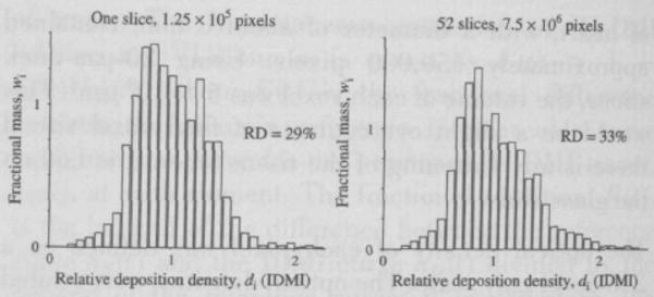
Distribution of relative deposition density for IDMI in the left ventricular myocardium of an open chest hamster. Left panel: One slice, providing 125,000 pixels of 16 × 16 μm, RD = 29%. Right panel: Aggregate distribution from the same heart using all 52 slices, for a total of 7.5 × 106 pixels of the same size.