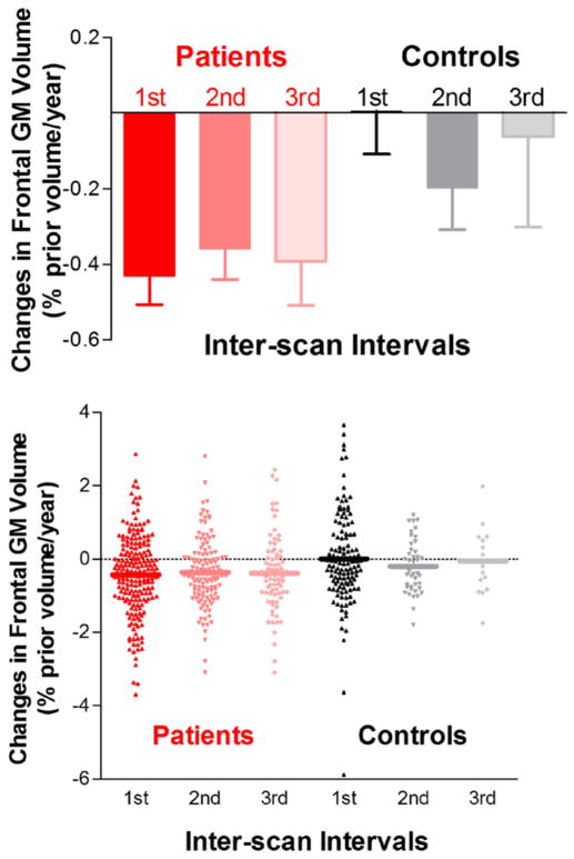
Figure 1.
Bar graphs and scatter plots illustrating the pattern of brain changes over time. Frontal gray matter (GM) changes in schizophrenia patients were most pronounced early in the course of schizophrenia. Frontal GM in schizophrenia patients differed significantly from healthy volunteers during the first interscan interval but not during subsequent intervals.

