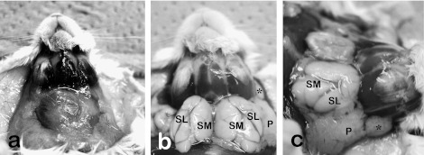
Fig. 1.
Macroscopic anatomy of the anterior and lateral neck portions of mice before (a) and after (b, c) the perfusion of fixative and removal of fat tissues. Anterior neck is occupied by large white fat tissues including salivary glands (a). Upon removing fat tissues and submandibular lymph nodes, salivary and extraorbital lacrimal glands were exposed. The extraorbital lacrimal gland (*) is located anterior to the parotid gland (P). The submandibular (SM) and sublingual (SL) glands are encapsulated with common fascia.

