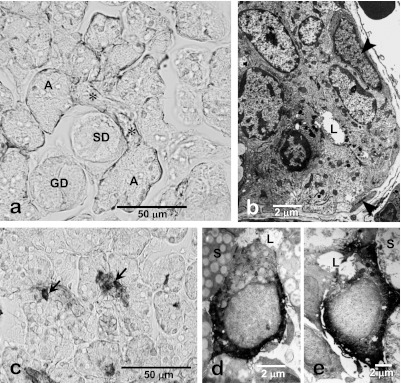
Fig. 3.
Immunohistochemistry and electron microscopy of the intercalated duct of the rat submandibular gland. Smooth muscle-action-immunohistochemistry shows distribution of myoepithelial cells along the intercalated duct (*) as well as acini (A). No myoepithelial cells are associated with other duct portions such as the striated (SD) and granular ducts (GD). The intercalated duct cells are cuboidal in shape and partially surrounded by myoepithelial cells (arrowhead, b). Hsp27-immunohistochemistry of 3-week-old rat submandibular gland shows the centrally-located immunopositive terminal tubule cells (arrows, d). Electron micrograms of Hsp27-immunohistochemistry of 4-week-old rat submandibular gland show that Hsp27-immunoreactive terminal tubule cells differentiated into immature acinar cells (d) and granular intercalated duct cells (e). L, lumen; S, secretory granules in acinar cells.

