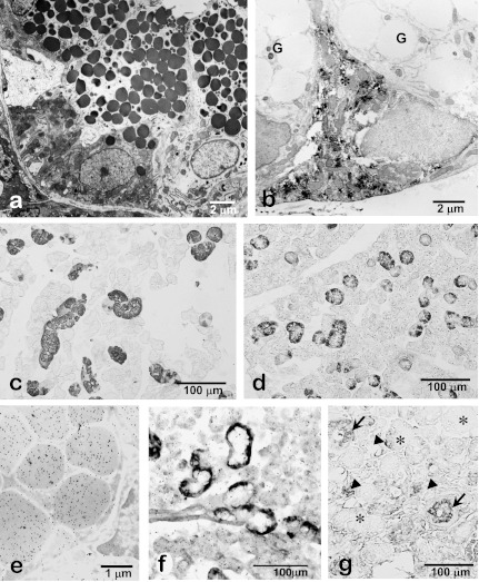
Fig. 4.
Photomicrographs of rat (a, b, e, f), mouse (c, d) and human (g) submandibular glands. Electron microscopy shows supranuclear localization of numerous exocrine secretory granules in the granular duct cells (a). FGF2-immunoelectron microscopy shows an immunopositive pillar cell (*) without large secretory granules present between the principal granular duct cells without FGF2-immunoreactivity (G) (b). EGF-immunohistochemistry of the submandibular glands shows specific localization of EGF in the granular duct in male (c) and female (d) mouse. Note that EGF-immunopositive granular ducts are less developed in the female submandibular glands than the male glands. Colloidal-gold particles representing the immunoreactivity for EGF are exclusively localized in the secretory granules of the granular duct cells (e). Expression of EGF-mRNA is specifically localized in the basal cytoplasm of the granular duct cells (f). In human submandibular glands, EGF-immunoreactivity is dominantly localized in the striated duct (arrows) and partially in the serous acinar cells (arrowheads). No immunoreaction is detected in mucous acini (*) (g).

