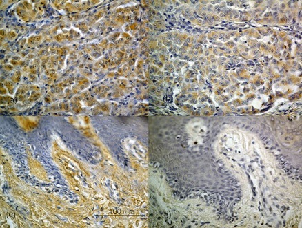
Fig. 2.
Immunohistochemical staining for CXCR4 and CXCL12 in malignant melanoma. Nodular melanomas showing a high CXCR4/CXCL12 immunoexpression percentage (a: CXCR4, b: CXCL12). The immunoexpression was detected diffusely in the cytoplasm. In most cases, the CXCL12 distribution was similar to that of CXCR4. However, in acral lentiginous melanomas, CXCR4/CXCL12 immunoexpression (c: CXCR4, d: CXCL12) was at very low percentage, and was partially observed in the cytoplasm of tumor cells (c, d). Original magnification ×400. Bar=100 µm.

