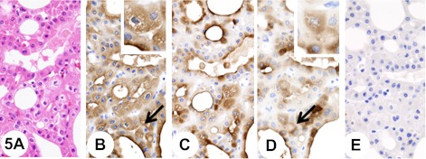
Fig. 5.
Chromophobe renal cell carcinoma, eosinophilic variant. Insets show higher-magnification views of the regions indicated by arrows. The tumor is mainly composed of tumor cells that are smaller and markedly eosinophilic, and often showed a perinuclear halo (A). The tumor cells show diffuse fine granular cytoplasmic staining reaction with anti-KL-6 antibody (B). In addition, immunostaining is seen at the luminal surface of tubules (B). Pretreatment with neuraminidase decreases immunoreactivity with anti-KL-6 antibody (C). The tumor cells show diffuse fine granular cytoplasmic staining reaction with anti-sialyl MUC1 antibody (D). In addition, immunostaining is seen at the luminal surface of tubules (D). Pretreatment with neuraminidase abolishes immunoreactivity with anti-sialyl MUC1 antibody (E).

