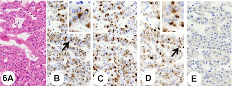
Fig. 6.
Renal oncocytoma. Insets show higher-magnification views of the regions indicated by arrows. The tumor is composed of large, well-differentiated, polygonal, eosinophilic cells with a cytoplasm that is rather granular. The tumor cells are arranged in solid nests and/or in cords separated by loose edematous stroma (A). The tumor cells show a polarized cytoplasmic staining reaction close to the nucleus with anti-KL-6 antibody (B). After the pretreatment with neuraminidase, the immunoreactivity with anti-KL-6 antibody remains (C). The tumor cells show a polarized cytoplasmic staining reaction close to the nucleus with anti-sialyl MUC1 antibody (D). Pretreatment with neuraminidase abolishes immunoreactivity with anti-sialyl MUC1 antibody (E).

