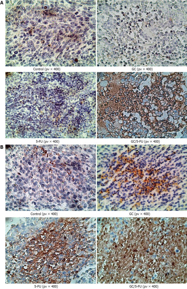
Figure 6.
Immunohistochemistry of p53 and caspase-3 in the tumor sections from mice with different treatments. A: p53 staining in the control and galactosylated chitosan (GC) groups showed a scattered nuclear distribution pattern, in dark yellow or dark brown; while in 5-fluorouracil (5-FU) and GC/5-FU groups, p53 showed a sheet staining pattern, which was more dramatic; B: Caspase-3 staining in the control and GC groups showed a scattered cytoplasmic distribution pattern, in dark yellow or dark brown; while in 5-FU and GC/5-FU groups, caspase-3 showed a sheet staining pattern, which was more dramatic in the GC/5-FU group.

