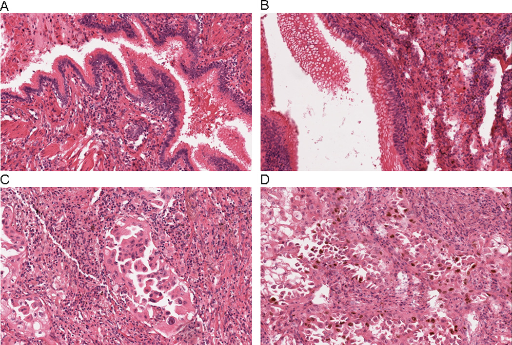
Figure 2.

Immunohistochemical localization of Ki67 in A) normal bronchial epithelium, placebo group, B) normal bronchial epithelium, β-carotene group, C) lung tumor cells, placebo group, and D) lung tumor cells, β-carotene group. In Panels A and B, nuclear positivity for Ki67 is essentially absent in columnar ciliated bronchiolar-type epithelium. In Panel C, only scattered malignant cells show nuclear staining for Ki67, whereas numerous positive-staining nuclei are apparent in Panel D. Rare scattered inflammatory cells are also positive for Ki67 in Panel D. All photomicrographs are 20× magnification.
