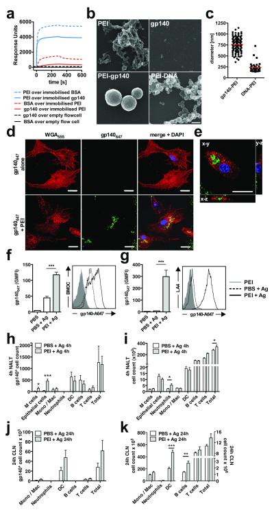
Figure 2.
Interactions between antigen and PEI and between immune cells and PEI-antigen complexes. (a) Direct biophysical interaction between PEI and BSA or PEI and gp140 analyzed using SPR with surface-immobilized protein or PEI. Data are representative of 3 independent experiments. (b) Scanning electron micrographs of PEI alone, gp140 alone or PEI complexed with gp140 or DNA; bar = 500 nm. (c) PEI-antigen complex formation. Quantification of PEI-gp140 or PEI-DNA particle sizes determined by counting of randomly-chosen fields imaged by SEM. (d) BMDC uptake of gp140647-PEI complexes. gp140647 (2 μg/mL) complexed or not with 0.1 μg/mL PEI. Cells were surface stained with Alexa Fluor® 555-labelled wheat germ agglutinin (WGA555) and nuclear stained with Hoechst 34580. Laser scanning confocal micrographs of BMDC incubated with gp140647 alone (top panel) or gp140647 mixed with PEI (bottom panel), bar = 20 μm; (e) 3D reconstructions of a single BMDC exposed to gp140647 complexed with PEI, bar = 10 μm. (f) Quantification of gp140647 uptake by BMDC or (g) LA-4 mouse lung epithelial cells after O/N exposure to antigen with or without PEI. (h-k) Uptake of gp140647 into NALT (h) or CLN (j) cells, or cell recruitment into NALT (i) or CLN (k), 4h (h, i) or 24h (j, k) after intranasal immunization of BALB/c mice with 10 μg gp140647 ± 20 μg PEI (n=5, two-tailed t-test). Leukocyte populations were identified by flow cytometry with appropriate labeling and gates. Data are representative of 2 or more independent experiments and are presented as mean values of replicates from one experiment ± SEM; * p<0.05, ** p<0.01, *** p=0.001

