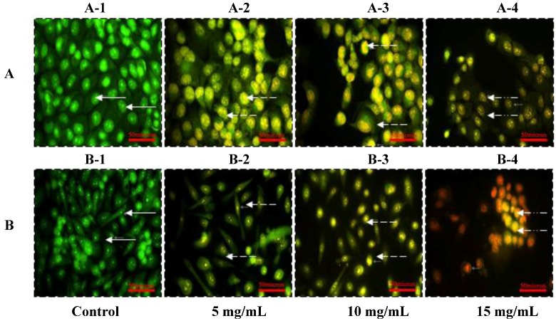
Figure 2.
Morphologic observation with acridine orange/ethidium bromide (AO/EB) staining. DU-145 cells (A) were treated without (A-1) and with SIO at 5 mg/mL (A-2), 10 mg/mL (A-3), and 15mg/mL (A-4) for 24 h. PC-3 cells (B) were treated without (B-1) and with SIO at 5 mg/mL (B-2), 10 mg/mL (B-3), and 15 mg/mL (B-4) for 24 h. ( ) indicates viable cells; (
 ) indicates early apoptotic cells; (
 ) indicates late apoptotic cells. Each experiment was performed in triplicate (n = 3) and generated similar morphologic features. Original magnification 400×, bar = 50 μm.

