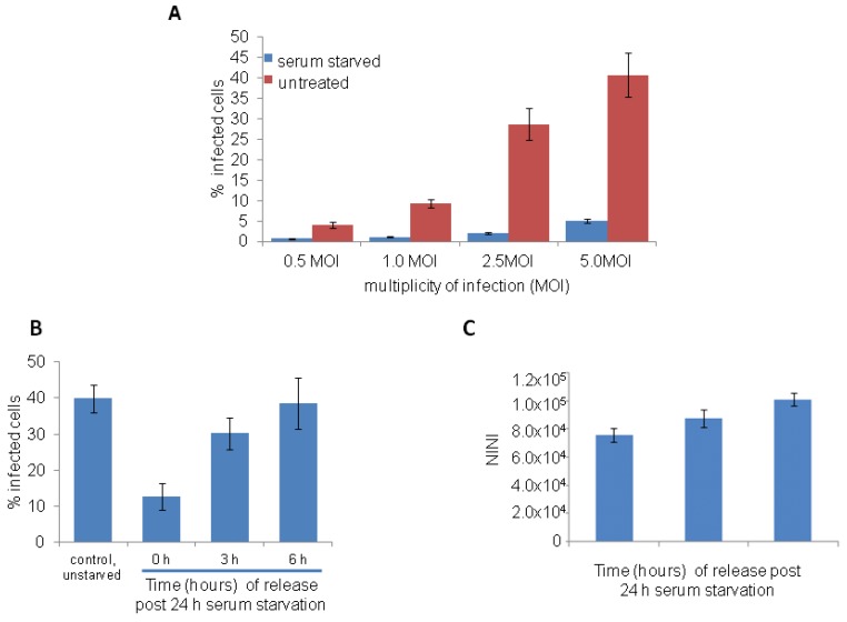
Figure 2.
Ebola virus (EBOV) infection is restricted in serum starved HeLa cells. (A) HeLa cells were serum starved for 24 h and then infected with different MOI of EBOV for 24 h in complete medium. Cells were stained with anti-GP antibody (6D8) and imaged. Image analysis showed reduced EBOV infection in serum starved cells compared to control cells. (B,C) HeLa cells were serum starved for 24 h and then infected at 0, 3 h and 6 h following release from the block with 5 MOI of EBOV. After 24 h, cells were fixed, stained with anti-GP antibody (6D8) and Hoechst 33342 dye, and acquired images were analyzed. A time-dependent increase in the EBOV infection (B) and increase in NINI values based on Hoechst 33342 dye (C) was observed.

