Figure 3.
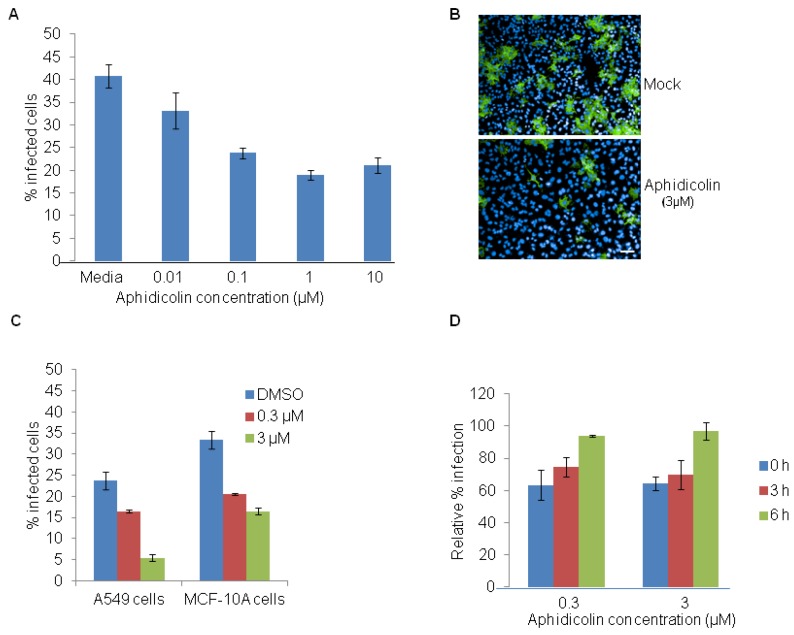
Aphidicolin restricts EBOV infection. (A) HeLa cells were pretreated with DMSO control or with indicated concentrations of aphidicolin for 12 h and then infected with 5 MOI of EBOV for 24 h in the absence of the inhibitor. A dose-dependent decrease in percentage of EBOV infected cells was observed. (B) Representative images of HeLa cells infected with EBOV and stained with anti-GP antibody (6D8) (green) and nuclear Hoechst dye (blue), following pretreatment with DMSO (top panel) or aphidicolin (3 μM, bottom panel). (C) A549 or MCF10A cells were pretreated for 12 h with DMSO control or with 0.3 μM or 3 µM aphidicolin and then infected with 5 MOI of EBOV for 24 h in the absence the inhibitor. A dose-dependent decrease in percentage of EBOV infected cells was observed. (D) HeLa cells were released from aphidicolin block and infected with EBOV at 0, 3 or 6 h post-release. Results show a time-dependent increase in efficiency of EBOV infection. Scale bar, 20 µm.

