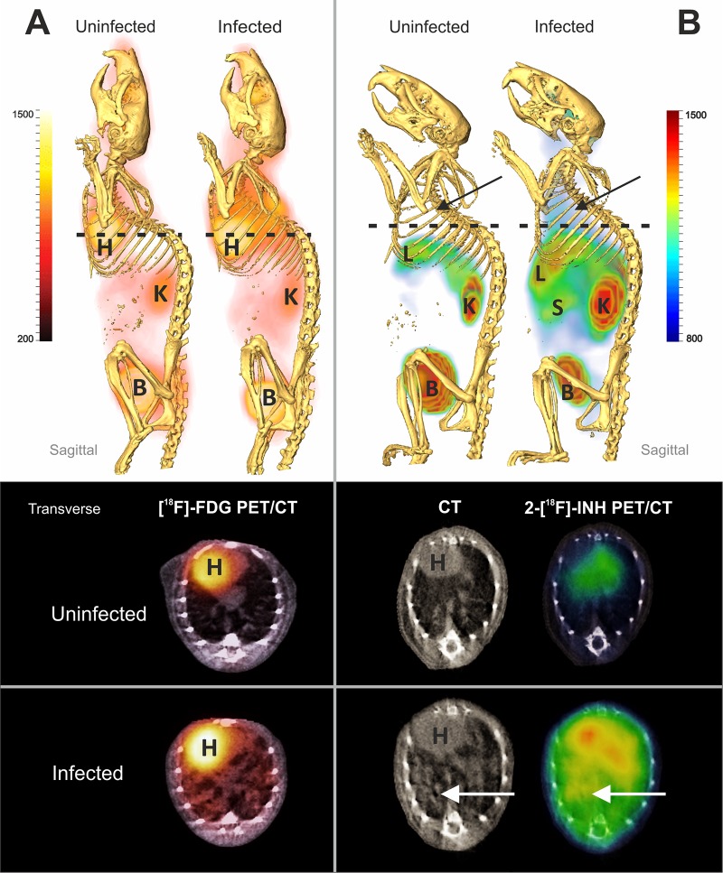
Fig 2.
PET/CT imaging of M. tuberculosis-infected mice with diffuse pulmonary disease. Infected and uninfected BALB/c pairs were imaged after injection of 2-[18F]-FDG (A) or 2-[18F]-INH (B). Whole-animal sagittal and transverse sections are displayed as combined PET/CT images with heart (H), liver (L), kidneys (K), spleen (S), and bladder (B) marked. [18F]FDG-PET activity is noted in the lung fields in infected animals but not uninfected animals (A). Background uptake is noted in the heart, kidneys, and bladder of both the uninfected and infected animals. Similarly, diffuse 2-[18F]-INH-PET activity is noted in the lung fields of the infected animal but not the uninfected mouse (B). Background PET signal in the heart, liver, kidneys, and bladder is noted in both infected and uninfected animals.

