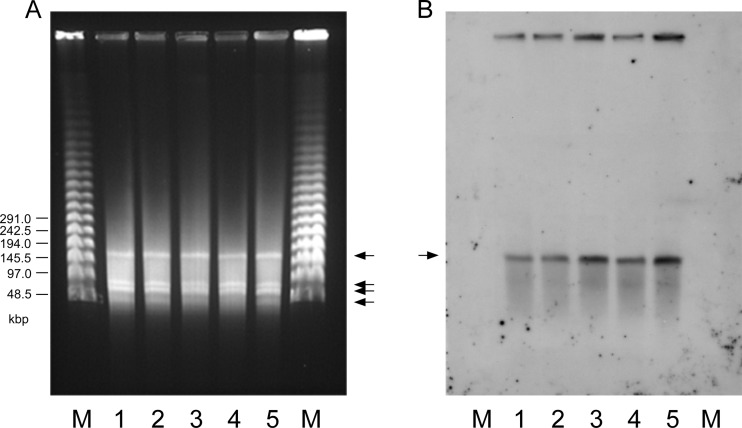
Fig 2.
PFGE analysis of the plasmid DNAs of VanN-type GRE isolates using S1 nuclease enzyme and Southern hybridization with the vanN ligase gene probe. (A) PFGE analysis using S1 nuclease enzyme. At least four plasmid DNA bands were detected on the gel (arrows). (B) Southern hybridization of panel A blotted with the vanN ligase gene labeled using a nonisotopic digoxigenin system (Roche). Of the four plasmid bands, the largest, a 160-kbp plasmid DNA band, bound to the vanN-specific probe (arrow). Lanes M, lambda ladder PFGE molecular size marker (NEB); lanes 1, E. faecium GU121-1; lanes 2, GU121-2; lanes 3, GU121-3; lanes 4, GU121-4; lanes 5, GU121-5.

