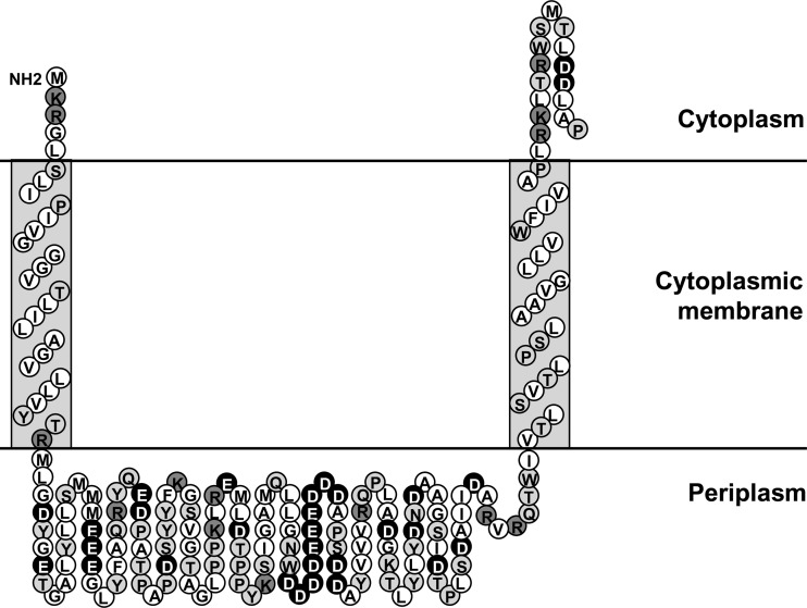
Fig 1.
Topology prediction of the N-terminal portion (amino acids 1 to 187) of the sensor kinase CprS, showing the two transmembrane helices and the periplasmic sensor domain. Hydrophobic residues are represented by the white circles, polar residues are represented by the light-gray circles, and positively and negatively charged residues are represented by the dark-gray and black circles, respectively. This figure was developed based on the output of the program Sosui 1.11 (17).

