Figure 1.
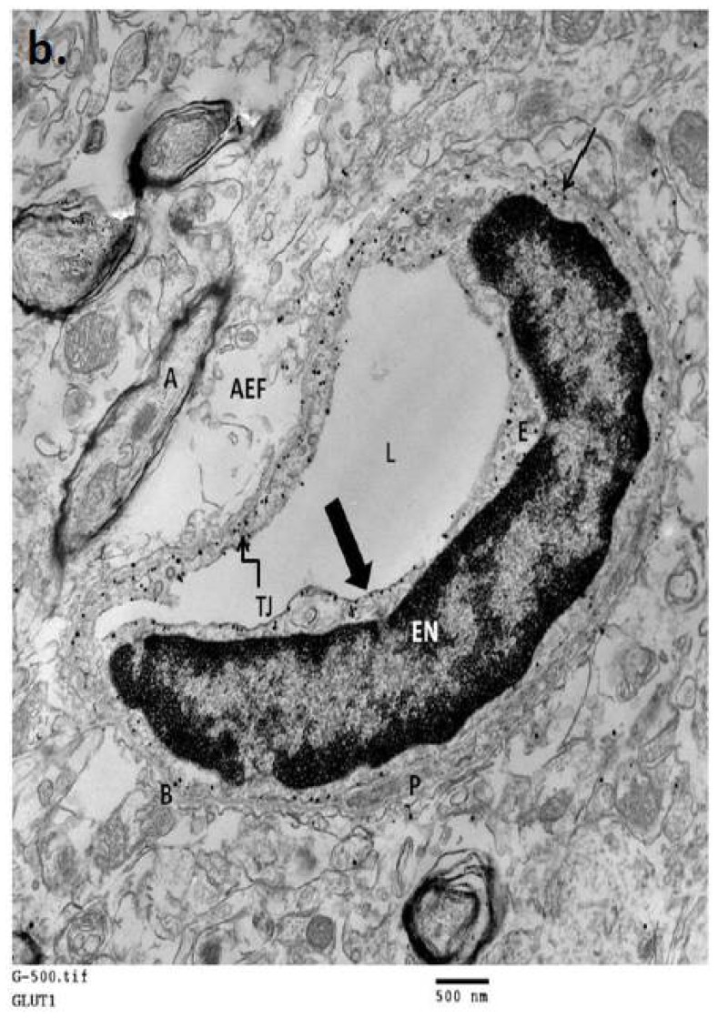
(a) A schematic representation of the cellular localization of glucose transporters (GLUTs and SGLTs) in mammalian neurovascular unit; (b) An electron micrograph of normal mouse brain neurovascular unit showing polarized expression of GLUT1 transporter in endothelial cells. L-lumen (blood side); E-endothelial cell; EN-endothelial nucleus; P–pericyte; B-basement membrane; TJ-tight junction; AEF-astrocyte end foot; A-myelinated axon; Thick arrow, shows luminal membrane with 6 nm silver enhanced immunogold labeled GLUT1 protein; Thin arrow, shows abluminal membrane with immunogold labeled GLUT1 protein. (Immunogold labeling and micrograph generated in the Abbruscato Lab.)


