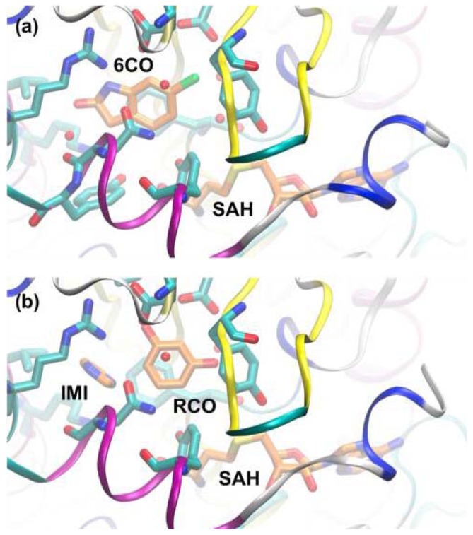
Figure 8.
(a) PNMT with 6-chlorooxindole (6CO) and S-adenosyl-homocysteine (SAH) modelled in the active site (based on PDB entry 3KPY); (b) Same structure after reassignment of density to imidazole (IMI) and resorcinol (RCO) (based on PDB entry 4DM3). Protein backbone is shown in ribbon form, with residues shown in stick form. Ligands are drawn in stick form with carbon atoms colored orange.

