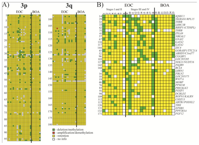
Figure 2.
Hybridization pattern of DNA from Epithelial ovarian cancer (EOC) and benign ovarian adenomas (BOA) samples on NotI-microarrays. (A) Vertically, 180 NotI sites arranged according to their localization on chromosome 3 (from 3p26.2 to 3p11.1 and from 3q11.2 to 3q29). Horizontally, 25 ovarian samples (18 EOC and 7 BOA); (B) Vertically, 35 NotI sites arranged by methylation/deletion frequency (from 33% to 17%).

