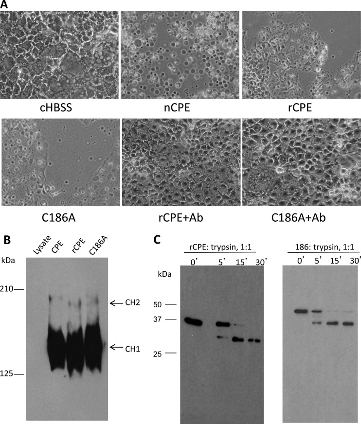
Fig 1.
Comparison of biochemical characteristics of the C186A variant versus rCPE. (A) Cellular morphological damage. To initially assess the cytotoxic properties of the C186A variant, a morphological-damage assay (33) was performed. Confluent Caco-2 cell monolayers were treated at 37°C for 60 min with HBSS or HBSS containing 2.5 μg/ml of nCPE, rCPE, or the C186A variant. After this treatment, Caco-2 cells were photomicrographed at a total magnification of ×400. To test the specificity of cytotoxicity caused by these toxins, the C186A variant or rCPE was preincubated at 37°C with CPE polyclonal antiserum for 15 min; those mixtures were then applied to confluent Caco-2 cells in 6-well plates. Shown are representative results from three repetitions of this experiment. Ab, antibody. (B) Large-complex formation by the C186A variant and rCPE. Confluent Caco-2 cells were treated with 2.5 μg/ml of either rCPE species for 60 min at 37°C. The treated cells were then loaded onto a 4.25% polyacrylamide gel containing SDS. Large-complex formation was evaluated by Western blotting of the gel using rabbit polyclonal anti-CPE serum. “CH1” and “CH2” represent the two CPE large complexes, which migrate anomalously as ∼155- and 200-kDa proteins, respectively, on these gels (26). (C) Limited trypsin digestion assay. To assess gross conformational changes in the C186A variant, both rCPE and the C186A variant (500 ng) were incubated with trypsin (2 μg) at 25°C for the periods indicated above the lanes. After samples were stopped by the addition of Laemmli buffer and boiling for 5 min, proteins in those samples were separated by electrophoresis on an SDS-containing 10% polyacrylamide gel, followed by Western blotting with rabbit anti-CPE polyclonal antibody.

