Fig 2.
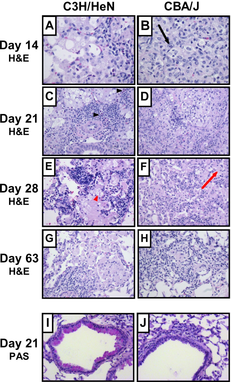
Lung histology following C. neoformans infection. Inbred 7-week-old female C3H/HeN and CBA/J mice were intratracheally infected with 104 CFU of C. neoformans ATCC 24067, and lungs were fixed, excised, paraffin embedded, and stained with H&E (A to H) or PAS (I and J) at the indicated time points. Photomicrographs of representative lung sections from 2 to 4 mice were taken at a magnification of ×200. Airway epithelial mucus stains red with PAS; highlighted cells include C. neoformans (red arrowhead), macrophage (red arrow), neutrophil (black arrow), and eosinophils (black arrowheads).

