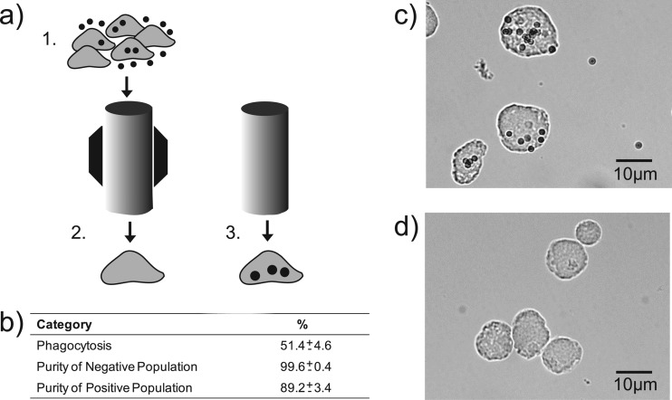
Fig 1.
Sorting of phagocytic and nonphagocytic E. histolytica trophozoites. (a) Diagram depicting the steps of the magnet-assisted sorting of amoebic subpopulations where E. histolytica were allowed to phagocytose C1q-coated paramagnetic beads (step 1), applied and washed through a magnetized column whereby the negative population is eluted (step 2), and then the magnet is removed and the positive population is eluted (step 3). (b) Table showing percent phagocytosis and the purity of eluted populations based on manual cell counts (mean and standard error, n = 5). Photomicrographs show ×40 representative images of both the positive (c) and negative (d) amoebic populations.

